Clinical Features of AGA in Women
Androgenetic Alopecia (AGA) also known as female pattern hair loss in women is a genetic condition and is caused by androgens in both men and women. The incidence of this condition is the same for men as it is for women. It beings between the ages of 12 and 40 years and is inherited in a polygenic manner. The hair follicles that are susceptible to this condition, the dihydrotestosterone will bind itself to the androgen receptor and then the hormone-receptor complex leads to activating the genes that are ultimately responsible for a gradual change to miniaturized follicles from large terminal follicles.
Levels of 5x reductase and androgen receptors are higher in frontal hair follicles when compared to occipital follicles in both young men and women. Men who have minimal aromatase have lower levels of cytochrome p-450 as compared to young women in the frontal follicles, whereas women have higher levels of aromatase in their occipital follicles. AGA is detected in women at an early age when the pattern of increase thinning over the occipital scalp presents itself with retention of the frontal hairline with a presence of miniaturized hair. Women who suffer from AGA do not have any problems with pregnancies and menses. Symptoms such as severe unresponsive cystic acne, galactorrhea, civilization, and hirsutism should be paid attention to, and no hormone tests need to take unless these symptoms show up. Women with AGA can make use of a Topical minoxidil solution as it is the only drug that will promote hair growth. The drug has been proven by double-blind studies that have used hair weight and hair counts.
Pathophysiology
AGA, also known as female pattern hair loss is a distressing and common issue faced by both men and women. Even though it is very common many clinical investigators and clinicians have trouble diagnosing it in women. It may begin between the ages of 12 and 40 years in both men as well as women, and 50% of the population is known to experience this condition before they hit the age of 50. This condition could be inherited from either side of the family.
This condition is genetically induced by androgens in women and in men. Men and women who suffer from this condition have high levels of 5x-reductase and dihydrotestosterone (DHT), formed by the peripheral conversion of testosterone, and is also responsible for the miniaturization of scalp hair follicles. In follicles that are genetically susceptible, DHT will bind itself to the androgen receptor as well as to the hormone-receptor complex, thus activating the genes that are responsible for the slow transformation of large terminal follicles into small, miniaturized follicles. With time and with successive AGA cycles, anagen duration becomes shorter, the matrix size becomes less which leads to smaller follicles that produce finer, shorter, and miniaturized hair that do not cover too much of the scalp area. Miniaturized hair of different lengths is characteristic to AGA, while the number of follicles per unit area remains the same.
Men experience more hair loss with AGA as compared to women. The hormonal basis for the basis is the same for men as well as women. To confirm the hormonal basis, a study was conducted in 12 young men and women. Type 1 and Type II 5x-reductase, androgen receptor and cytochrome p-450 aromatase were measured in their hair follicles through scalp biopsies.
Both men and women showed higher levels of Type 1 and Type II 5x-reductase and androgen receptors in the frontal hair follicles compared to occipital hair follicles. However, these levels were half as compared to those of men. Young women had high levels of cytochrome p-450 aromatase in the frontal follicles as compared with men who had hardly any aromatase, women, on the other hand, has much higher aromatase levels in the occipital follicles. One should note the differences in the aromatase that can convert testosterone to estradiol. The study also suggests that a milder version of AGA may appear in women because of lower levels of 5x-reductase and androgen receptors present in the frontal follicles as compared to the present in men. High levels of aromatase levels in women can cause an increase in the formation of estradiol from testosterone and less 5x-reductase products like DHT.
Even though the hair thinning pattern for women may diffuse, there exists a pattern for both men as well as women, where there is higher hair density in the parietal and frontal scalp. If you look at it from a clinical perspective you will note that hair follicles on the frontal and parietal scalp differ from follicles on the occipital scalp. In AGA, occipital follicles are usually spared by the influence of hormones, whereas occipital follicles affected by the ophiasis pattern are more resistant to regrowth. The embryological derivation of the dermis in the occipital and frontal/parietal regions causes the difference in behaviors of the follicles. Avian embryology suggests that the dermis of the parietal/frontal scalp has a neural crest origin. Cell-cell interaction, regulatory genes, and growth factors influence the hair follicles to affect the two regions very differently depending on where their dermal origin is, and this is what causes different patterns in scalp hair loss.
In most cases, women will notice some thinning of hair in the frontal area and then the scalp starts becoming more visible. With time, even though the thinning may be diffuse, it may take over most of the scalp. It will usually pattern itself with marked thinning over the frontal and parietal scalp and more density over the occipital scalp. Women will usually retain a think rim of hair along the frontal hairline, and in most cases, the scalp will be seen behind the hairline. Shortening of the Anagen phase and reduction of the matrix size cause miniaturized hair, shorter hair of different diameters and lengths to appear, which is also characteristic of AGA. Since there is increased space between the hairs, it will make the central part look wider over the frontal part compared to the occipital area.
Sometimes, hair density may appear normal, but hair does not grow back to its normal length, which leads to wispy distal ends. This happens when the shortening of anagen happens faster than matrix reduction. The patient will note that the ponytail is no longer as thick and hair would have to be cut to shorter lengths to make the appearance look fuller. Advanced thinning rarely occurs with the loss of the frontal hairline, but if it does, it is associated with elevated circulating androgens.
In women, AGA is not accompanied by an increase in shedding. But, in some cases like telogen effluvium which is followed by childbirth, any major illness or some other cause may make one susceptible to it. Women who do not comb their hair after shampooing and let them dry naturally may not know exactly if their hair is shedding. They will find strands of hair in the hands when they comb their hands through the semi-dry hair.
Laboratory Evaluation
In most cases, one may not need to go in for extensive hormone testing unless there are signs that there is excess androgen present. People who have AGA have normal pregnancies and menses. Women are asked to undergo an endocrine evaluation only after they have been identified after asking them questions about their history of fertility, about their menses, severe unresponsive cystic acne, virilization, presence of hirsutism, and galactorrhea. If even one of these is present, then laboratory measurement of serum total, free testosterone, prolactin, and dehydroepiandrosterone sulfate are indicated. Other causes can be ruled out by the measurement of serum thyrotropin, and iron studies that include serum iron and ferritin, RPR, and complete blood count.
Differential Diagnosis
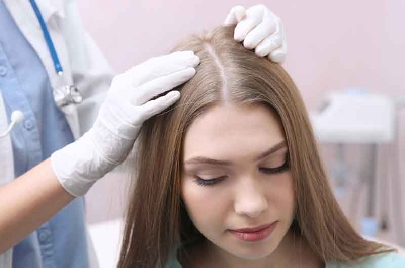
AGA is diagnosed in women when it comes in at an early age, there is greater thinning over the frontal/parietal scalp with more hair density around the occipital scalp. There is the retention of the frontal hairline and miniaturized hairs are present.
These features make diagnosing AGA in women easy. Even though AGA is a common condition, other reasons for hair loss too need to be considered, recognized or even excluded. A scalp biopsy will help achieve this effect. Biopsies are usually taken from representative active sites in horizontal sections as this allows a larger number of follicular structures to be collected and studied. In case an AGA is detected, it will show an increase in the number of miniaturized hairs, abundant and enlarged sebaceous glands as well as minimal inflammation.
CTE or Chronic Telegon Effluvium may come up with a diagnosis that is difficult and differential, even though the feature may be distinct. Women between the ages of 40 and 60 with CTE, who have above average hair density, may mark a sudden onset of heavy shedding on their entire scalp. Even though the shedding may be prolonged, the hair will appear of normal density or minimally decreased, a hair pull test will extract telogen hairs easily. Miniaturized hair is not seen. Horizontal sections in a scalp biopsy help distinguish between a CTE and AGA. The ratio of miniaturized hair to terminal hairs in CTE is 1:9 and in AGA is 1:2 and for a normal scalp is 1:7.
Acute Telogen effluvium may occur in a woman who has a long-standing condition of unmasked latent AGA or AGA. Learning the history of the patient to understand the cause of profuse hair shedding can reveal conditions like severe dietary protein deficiency, high fever, or chronic blood loss caused in women due to heavy menses. There is a category of drugs that can cause hair loss and they include anticoagulants, anticancer drugs, antithyroid drugs, anticonvulsants, beta-blockers, tricyclic antidepressants, and progestins that have androgenic effects.
Diffuse and patchy Alopecia and may coexist with AGA and in some cases could present a differential that is challenging and can only be resolved by a scalp biopsy that shows a peribulbar lymphocytic infiltrate around the anagen hair bulbs. An adult who has loose anagen syndrome will have a history that involves decreased hair density since early childhood and the pull test will be true. If the extracted hairs were examined under a low power microscope, misshapen anagen bulbs, a ruffled cuticle, a nonexistent inner root sheath.
Medical Treatment of AGA in Women
The only topical drug that is approved to treat AGA in women is Topical minoxidil. Its efficacy has been proven in placebo-controlled, double-blind studies with the help of hair counts and hair weights as the primary endpoints. Women aged between 22 – 41 who used a 2 % Minoxidil solution experienced higher hair count and an increase in hair weight, compared to those that were applied a placebo in 16 weeks. Clinical perception of improved scalp coverage may take up to 6 – 12 months. This solution needs to be applied at least twice a day to dry scalp.
Finasteride is an inhibitor of type II 5x-reductase and is contraindicated in women who may become or may be pregnant because these could cause trouble to the external genitalia in the male fetus. It did not work for postmenopausal women in a study that was controlled by a placebo.
Women are disappointed with thinning hair and their hair needs to be evaluated and managed and also need some reassurance that they can safely use cosmetics that will make their female pattern baldness hairstyles look more fuller.
References
Cash TF, Price VH, Savin RC, Psychological effects of androgenetic alopecia on women. Comparisons with balding men and with female control subjects. J Am Acad Dermatol 29:568-575, 1993
Dallob AL, Sadick NS, Unger W, et al: The effect of Finasteride, a 5x-reductase, inhibitor, on scalp skin testosterone and dihydrotestosterone concentrations in patients with male pattern baldness. J Clin Endocrinol Metab 79:703-706, 1994
Frieden IJ, Price VH: Androgenetic alopecia. In: Theirs BH, Dobson RL (eds). Pathogenesis of Skin Disease. New York: Churchill Livingstone, 1986: p 41-55
Hamilton JB: Patterned loss of hair in man: Types and incidence. Ann N Y Acad Sci 53:708-728, 1951
Headington JT: Transverse microscopic anatomy of the human scalp. Arch Dermatol 14:449-456, 1984
Kaufman K: Androgen metabolism as it affects hair growth in androgenetic alopecia. Dermatol Clin 14:697-711, 1996
Kuster W, Happle R: The inheritance of common baldness: Two B or not two B? J Am Acad Dermatol 11:921-926, 1984
Lattanand A, Johnson WC: Male pattern alopecia. A histopathologic and histochemical study. J Cutan Pathol 2:58-70, 1975
Ludwig E: Classification of the types of androgenetic alopecia (common baldness) occurring in the female sex. Br J Dermatol 97:247-254, 1977
Messenger AG: The control of hair growth: An overview. J Invest Dermatol 101, (1 Suppl): 4S-9S, 1993
Mirmirani P, Price VH: Top 10 Misconceptions about androgenetic alopecia in women. Cosmetic Dermatology 30-31 December 2000
Olsen EA: Androgenetic alopecia. In: Olsen EA (ed). Disorders of Hair Growth: Diagnosis and Treatment. New York: McGraw-Hill, 1994; p 257-283
Price VH: Testosterone metabolism in the skin. A review of its function in androgenetic alopecia, acne vulgaris, and idiopathic hirsutism including recent studies with antiandrogens. Arch Dermatol 111:1496-1502, 1975
Price VH: Treatment of Hair Loss. N Engl J Med 341:964-973, 1999
Price VH, Gummer CL: Loose anagen syndrome. J Am Acad Dermatol 20:249-256, 1989
Price VH, Menefee E: Quantitative estimation of hair growth I. androgenetic alopecia in women: effect of minoxidil. J Invest Dermatol 95:683-687, 1990
Price VH, Roberts JL, Hordinsky M, et al: Lack of efficacy of finasteride in postmenopausal women with androgenetic alopecia. J Am Acad Dermatol 43:768-776, 2000
Sawaya ME, Price VH: Different levels of 5x-reductase type I and II, aromatase, and androgen receptor in hair follicles of women and men with androgenetic alopecia. J Invest Dermatol 109:296-300, 1997
Schweikert HU, Wilson JD: Regulation of human hair growth by steroid hormones. I. Testosterone metabolism in isolated hairs. J Clin Endocrinol Metab 38:811-819, 1974
Sreekumer GP, Pardinas J, Wong CQ, et al: Serum androgens and genetic linkage. Analysis in early-onset androgenetic alopecia. J Invest Dermatol 113:277-279, 1999
Trancik RJ, Spindler JR, Cuddihy RV, et al: Clinician survey evaluating minoxidil topical solution in the treatment of androgenetic alopecia in patients under 18 years of age. Poster presented at 3rd Intercontinental Meeting of the Hair Research Societies, June 13-15, 2001: Tokyo, Japan, p129
Uno H, Cappas A, Schlagel C: Cyclic dynamics of hair follicles and the effect of minoxidil on the bald scalps of the stump-tailed macaques. Am J Dermatopathol 7:283-297, 1985
Venning VA, Dawber RPR: Patterned androgenetic alopecia in women. J Am Acad Dermatol 8:1073-1077, 1988
Whiting DA: Male pattern hair loss: Current understanding. Int J Dermatol 37:561-566, 1998
Whiting DA: Chronic telogen effluvium: Increased scalp hair shedding in middle-aged women. J Am Acad Dermatol 35:899-906, 1996
Whiting DA: Diagnostic and predictive value of horizontal sections of scalp biopsy. Specimens in male-pattern androgenetic alopecia. J Am Acad Dermatol 28:755-763, 1993
Whiting DA: The value of horizontal sections of scalp biopsies. J Cutan Aging Cosmet Dermatol 1:165-173, 1990
Ziller C: Pattern formation in neural crest derivatives. In: Dvan Neste, Randall VA (eds). Hair Research for the Next Millenium. Amsterdam, The Netherlands: Elsevier Science B, 1996: p 19-23